Posts

Showing posts with the label Cell Biology

Microbodies

These are  single  layered membranous sac like structures formed from the vesicles of endoplasmic reticulum or golgi bodies. These are - Peroxisomes   or Glyoxisomes .   Peroxisomes    Peroxisomes are formed from pre-existing peroxisomes. These are found in plants showing  “Photorespiration” (C -3 plants). Peroxisomes participate in the process of photorespiration along with choloplast and mitochondria.In peroxisome oxidation of glycolic acid (produced in chloroplast in photorespiration)  occurs.   These are also found in animal cells like “Protozoons” residing in liver or kidney cells. In peroxisomes ,catalases,oxidases enzymes are present. These enzymes assist degradation of toxic materials in cells. Peroxisomes invole in fat-metabolism in animal cells.   Glyoxisomes These are also single layered membrane bound spherical bodies found in cell showing “ Glyoxylate cycle ”  Enzymes of glyoxisomes participate in...

The Centrosome

Image
“Centrosome”term was first used by T.Boweri .  This is found mostly in animal cell and some algae.  Each centrosome  includes two pairs of “Centrioles”. Both pairs are placed at the angle of 90⁰ from one another. Centrioles of a pair are termed as “Diplosomes”.  A centriole is composed of nine triplet of microtubules. These triplets are arranged around a hub and embedded in amorphous dense substance.  A circle around centriole is called as centrosphere.RNA is also present in centrosome. Centrosome  participates in cell division.

Plasma membrane

Image
The membrane surrounding the cytoplasm of the cell is known as “ Plasma membrane or Plasmalemma ” .It is the outermost layer of animal cell while cell wall is present outside of plasmalemma in plant cell. Plsmalemma is a unit membrane and differentially permeable(permeable for some substances). Structure -  The structure of plasma membrane can be understood by “ Fluid-mosaic model ”  given by S.J.Singer & G.Nicolson in1972. According to this model in the middle of membrane bimolecular lipid layer is present. Peripheral proteins are present on outer surface of lipid layer while Intrinsic proteins are embedded in lipid layers. Peripheral proteins can be separated easily while intrinsic proteins cannot be separated easily. Peripheral proteins are glycoproteins and some glycolipids are also present with them. Glycoproteins and glycolipids are specific  with cells. Cells can recognize cell with same glycoproteins and glycolipid. This specification plays important r...

The Lysosome (Suicidal bag of the Cell)

Image
Lysosomes are single membrane bound structures found in animal cells. These were discovered by  D’Duve (Nobel prize-1974).    It is formed by golgi bodies or endoplasmic reticulum. Lysosome contains many digestive enzymes like “proteases,lipases,carbohydratases ,etc”. These become active at the acidic pH.   Many types of lysosomes are found as “Primary lysosomes,secondary lysosomes,residual bodies ,autophagic vacuoles”. Lysosome is considered as “ Suicidal bag of cell”  because its enzyme can digest all the parts of cell when lysosome bursts.   Function – ·      Primary lysosome  helps in digestion of material taken by cell through endocytosis. ·        Lysosomes help in autolysis of injured cells. ·        Lysosomal enzymes facilitates intracellular digestion. ·        Lysosomal enzymes digest food inside protozoans.

The Ribosome

Image
Ribosomes are minute structure associated with protein synthesis in the cell.  These are found in all cells, in mitochondria, chloroplast and nucleus of the cell.They are composed of RNA and proteins and not surrounded by any membrane. On the basis of “sedimentation coefficient”. Ribosomes are of two types- 70S ribosome –These are found in mitochondria ,chloroplast and prokaryotic cells like bacterial cell. It is composed of two units ,larger unit is 50S and smaller unit is 30 S. 80S ribosome -These are found in eukaryotic cells.  It’s larger unit is 60S and smaller unit is 40S. Ribosomes Mg + ion is required for union of two subunits of  ribosomes. Function- Ribosomes involves in protein synthesis.

The Nucleus

Image
Nucleus was discovered by “Robert Brown”. Nucleus has following parts -   Nuclear envelope - This is made of two parallel membrane .Space between membranes is known as  Perinuclear space . Nuclear envelope separates material inside nucleus from cytoplasm. In nuclear envelope some minute pores are present i.e nuclear pores. These acts as passage for RNAs and proteins between nucloplasm and cytoplasm. Nucleoplasm – Fluid inside nucleus is called as “Nucleoplasm”.   This contains “Nucleolus and Chromatins”.         Nucleolus – These are spherical bodies found in nucleoplasm. These were discovered by “Fontana”. Nucleolus is not a membrane bound structure. It is a site of ribosomal RNA synthesis. Numerous nucleoli are found in cells actively carrying out protein synthesis.       Chromosomes: These threads like structures are made of nucleoprotein  (DNA+Histone proteins)  i.e. chromatin. Chro...

The Golgi apparatus

Image
Golgi apparatus were first observed by Camillo Golgi (1898) . These are called "dictiyosomes" in plant cells. Golgi apparatus consists of many flat disc shaped sacs i.e. “Cisternae”. Cisternae are stacked parallel to each other. Golgi cisternae are concentrically arranged near the nucleus. Its forming face is convex i.e. “ cis” and maturing face is concave i.e. “ trans ” face. Both faces of golgi apparatus are different but interconnected. Functions – The golgi apparatus performs the functions of packaging materials and deliver either to intra-cellular targets or to secrete outside of the cell. Material to be packaged in the form of vesicles from the endoplasmic reticulum fuse with “cis” face and move towards “trans” face. Proteins formed by ribosomes on endoplasmic reticulum are modified in cisternae of golgi bodies before being released from “trans” face. Golgi apparatus are associated with the synthesis of glycoproteins and glycolipids. Golgi appa...

The Endoplasmic Reticulum

Image
 In eukaryotic cells,  a network of tubular structures are found in cytoplasm i.e. endoplasmic reticulum. Endoplasmic reticulum is found connected to the outer nuclear membrane. The endoplasmic reticulum are of two type. Rough Endoplasmic Reticulum -  This endoplasmic reticulum is attached to the outer nuclear membrane .Ribosomes are attached on the surface of these endoplasmic reticulum. RER are associated with protein synthesis and packaging. Smooth endoplasmic reticulum - These endoplasmic reticulum  are without ribosomes . These are associated with synthesis and storage of lipid. In the endoplasmic reticulum following three components are found- Cisternae - These are long ,flattened, unbranched and paralle arranged   tubules . Tubules –These are small ,smooth- walled branched tubular structures. Vesicles – These are rounded sac like structures. Sarcoplasmic reticulum is the modification of endoplasmic reticulum and found in the...

Plastids

Image
Plastids are particular feature of plant cell. They contains pigments and impart colours to the plants . On the basis of pigments, plastids are of following types-  Chloroplast –This plastid contains Chlorophyll. This traps photo-energy for photosynthesis.   Chromoplast – These are colour plastids other than chloroplast. Chromoplast  contain fat soluble pigments like carotenoids,xanthophylls etc.    Leucoplast - These are colourless plastid and store nutrients These are of following types.   Amyloplasts-store carbohydrates.   Elaioplasts-store oils and fats.   Aleuroplast-store proteins. Amyloplast of Potato Chromoplast in fruits and flowers Structure of chloroplast   Chloroplast is a double membrane bound cell  o rganelle like mitochondria. Space inside to the inner membrane of chloroplast is filled with fluid i.e. “Stroma” . In stroma many plate like membranous sacs are found and known as “Thylakoids...

Mitochondria (Power house of the cell)

Image
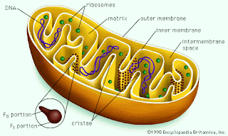
                                         Mitochondria are found in eukaryotic cells. Each mitochondrion is a double membrane bound structure. Space between two membranes is called “Outer mitochondrial space” while space inside to inner membrane is known as “Inner mitochondrial space”.  Fluid In inner mitochondrial space is called as “Matrix”. Inner mitochondria membrane forms many finger like  infoldings towards matrix .These infoldings are known as “Cristae” (singular –Crista). Cristae has many studded particles i.e. “F 1 particle or Oxysomes”.  Mitochondria are considered as “Semi-autonomous cell organelle because its matrix also contains circular DNA, some RNAs and 70S ribosomes. Mitochondria divide by fission. Structure of Mitochondria  Functions – Mitochondria are the site of aerobic cellular respiration.  It produce energy in the form ...

The Cell : Unit Of Life

Image
                                                                Body of livings are composed of many  tiny structures i.e. cell. Cell is considered as a structural, functional and biological unit of life .Cell is also known as building block of life.    Study of cell is called “Cytology”. The cell was discovered by “Robert Hook” in 1665. Cells in Onion peel   Cell theory  Schwann & Schleiden proposed "Cell theory" in 1839 . According to this theory- 1. All living things are made up of one or more cells. 2.Cells are the basic living units within organisms and the chemical reactions of of life take place within the cell. 3.All cells arises from pre-existing cells. Cells are of two types i.e. Prokaryotic cell and Eukaryotic Cell.      Prokary...